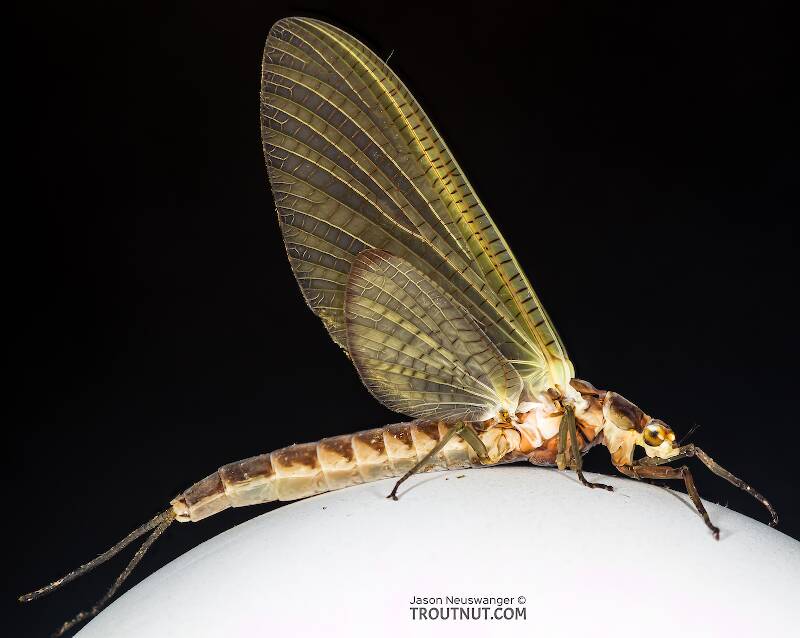
Hex Mayflies
Hexagenia limbata
The famous nocturnal Hex hatch of the Midwest (and a few other lucky locations) stirs to the surface mythically large brown trout that only touch streamers for the rest of the year.


Stonefly Species Soliperla quadrispinula (Roachflies)
Species Range
Identification
Species ID from GBIFthe Global Biodiversity Information Facility
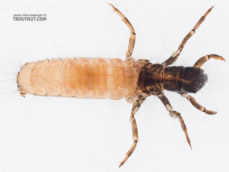
Based on these observations and supported by similar ones for Soliperla sierra and Soliperla thyra, we offer below a preliminary key to the larval Soliperla known from California.Source: California Soliperla Ricker, 1952 (Plecoptera: Peltoperlidae), Distribution And Taxonomic Characters
http: // lsid. speciesfile. org / urn: lsid: Plecoptera. speciesfile. org: TaxonName: 106 Figs. (2 - 3, 8 - 9, 14)
Physical description
Most physical descriptions on Troutnut are direct or slightly edited quotes from the original scientific sources describing or updating the species, although there may be errors in copying them to this website. Such descriptions aren't always definitive, because species often turn out to be more variable than the original describers observed. In some cases, only a single specimen was described! However, they are useful starting points.
Description from GBIFthe Global Biodiversity Information Facility
Source: California Soliperla Ricker, 1952 (Plecoptera: Peltoperlidae), Distribution And Taxonomic Characters
Male epiproct. Stark & Gustafson (2004) previously commented on variation in epiproct structure for this species. The SEM image in that study (Fig. 17 in Stark & Gustafson 2004), taken from a specimen collected in Elk River Canyon, Curry County, Oregon, shows the marginal armature consists of a single row of 19 irregularly spaced and sized teeth, with four additional submarginals and a width of 400 µm ; a small median gap occurs between teeth on the marginal row. The epiproct used in this study, taken from a Humboldt County, California specimen (Figs. 2 - 3) lacks most of the marginal teeth but the total number of teeth on the anterior face and margin (20) is similar to the number on the Elk River Canyon specimen, the width (417 µm) is also similar, and both samples show a median gap in the marginal teeth. Male aedeagus. Stark (1983) noted the total number of large setal spines, while often four, may be slightly more or less numerous due to extra or fewer spines on one or more aedeagal lobes. Three spines are visible on the specimen shown in Fig. 8, but the fourth is hidden. The largest visible spine (Fig. 9) is 300 µm long. Each spine originates from a long slender sclerite. Larval abdominal pigment pattern. Stark (1983) illustrated the pigment pattern for a specimen from the type locality in southern Oregon. This specimen had an obscure median circular spot on tergum 7 and relatively large median pale spots on terga 5 and 6. An additional small pale median spot is present on tergum 4. This is fairly similar to the pattern shown in Fig. 14 from a Humboldt County, California specimen. The median spot shown on tergum 6 for the latter specimen is smaller than the one on tergum 5, and it is more circular than triangular. Six additional Humboldt County specimens show similar patterns.
Start a Discussion of Soliperla quadrispinula
Stonefly Species Soliperla quadrispinula (Roachflies)
Species Range
Common Names
Resources
- NatureServe
- Integrated Taxonomic Information System
- Global Biodiversity Information Facility
- Described by Jewett, S.G. (1954) New stoneflies from California and Oregon (Plecoptera). The Pan-Pacific Entomologist 30, 167–180.

