Salmonflies
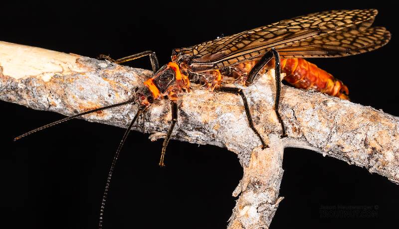
Pteronarcys californica
The giant Salmonflies of the Western mountains are legendary for their proclivity to elicit consistent dry-fly action and ferocious strikes.

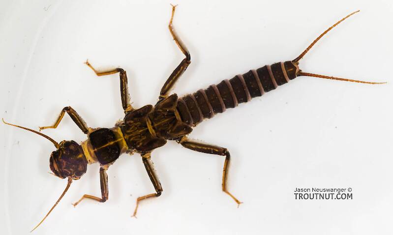
— 4 small yellow spots on frons visible in photos
— Narrow occipital spinule row curves forward (but doesn’t quite meet on stem of ecdysial suture, as it's supposed to in this species)
— Short spinules on anterior margin of front legs
— Short rposterior row of blunt spinules on abdominal tergae, rather than elongated spinules dorsally
I caught several of these mature nymphs in the fishless, tiny headwaters of a creek high in the Wenatchee Mountains.

Stonefly Species Soliperla sierra (Roachflies)
Species Range
Identification
Species ID from GBIFthe Global Biodiversity Information Facility
Male aedeagus. The structure and similarity of the aedeagus for this species to that of Soliperla campanula was documented by Stark (1983). Both species have a median row or grouping of relatively short, thick setal-spines on the ventral aedeagal surface (Fig. 10), and both have an anterolateral lobe with a close-set cluster of several additional short, thick setal spines (Fig. 11). The organization of the ventral setal-spine grouping for Soliperla sierra is not clearly in two well-organized rows, whereas in Soliperla campanula the two median rows are usually distinct. In addition, the anterolateral spine clusters are set on partially sclerotized lobes for mature specimens of Soliperla sierra, but these lobes are membranous for specimens of Soliperla campanula we have examined. Larval abdominal pigment pattern. The larva of Soliperla sierra is similar in body size and general features to those of known species. However, in the small sample available to us, the abdominal pigment pattern appears distinct from that of other Soliperla larvae known from California. Prominent, median longitudinal pale spots on terga 3, or 4 - 6 form an almost continuous median pale stripe on those segments. Tergum 7 often has a well defined circular median spot and the lateral pale spots on tergum 5 are much smaller than those on terga 2 - 4 (Fig. 15).Source: California Soliperla Ricker, 1952 (Plecoptera: Peltoperlidae), Distribution And Taxonomic Characters
http: // lsid. speciesfile. org / urn: lsid: Plecoptera. speciesfile. org: TaxonName: 105 Figs. (1, 4 - 5, 10 - 11, 15)
Physical description
Most physical descriptions on Troutnut are direct or slightly edited quotes from the original scientific sources describing or updating the species, although there may be errors in copying them to this website. Such descriptions aren't always definitive, because species often turn out to be more variable than the original describers observed. In some cases, only a single specimen was described! However, they are useful starting points.
Description from GBIFthe Global Biodiversity Information Facility
Source: California Soliperla Ricker, 1952 (Plecoptera: Peltoperlidae), Distribution And Taxonomic Characters
Male epiproct. Stark (1983) and Stark & Gustafson (2004) present images and brief descriptions of the Soliperla sierra epiproct based on a paratype specimen. The SEM image (Fig. 18 in Stark & Gustafson) shows an almost quadrangular shape, a wide toothless median gap along the marginal tooth row and 14 total teeth including 7 marginals and 7 submarginals. The anterior face of this specimen is 215 µm wide. The specimen examined in this study from Sierra County, California is oriented in an oblique fashion that emphasizes the width and the large toothless median gap. The SEM images of this specimen (Figs. 4 - 5) indicate 15 total teeth and a width of 268 µm.
Start a Discussion of Soliperla sierra
Stonefly Species Soliperla sierra (Roachflies)
Species Range
Common Names
Resources
- NatureServe
- Integrated Taxonomic Information System
- Global Biodiversity Information Facility
- Described by Stark, B.P. (1983) A review of the genus Soliperla (Plecoptera: Peltoperlidae). The Great Basin Naturalist 43, 30–44.

