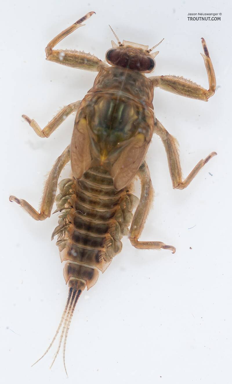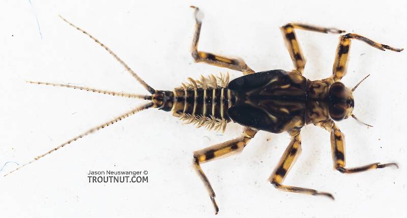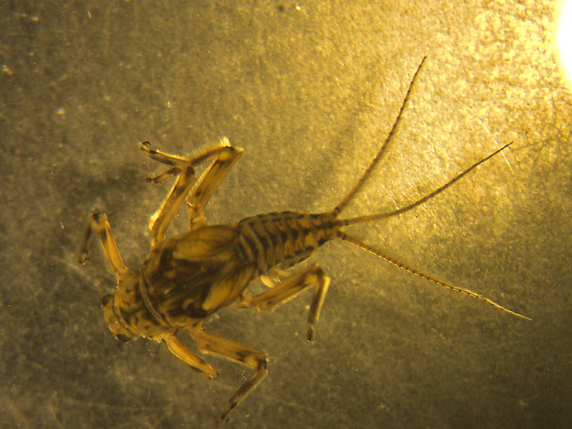
Salmonflies
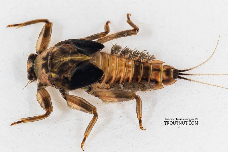
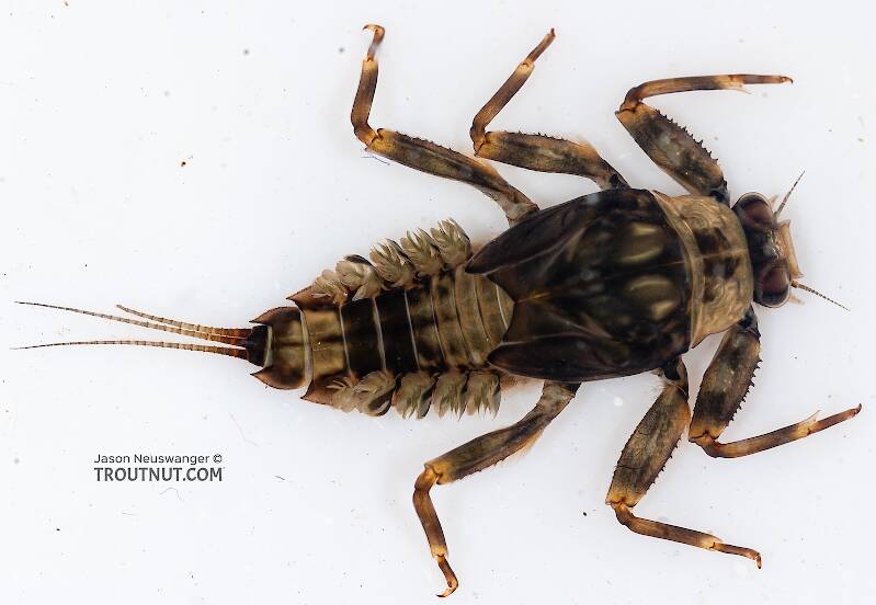
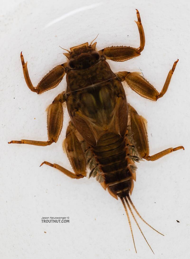
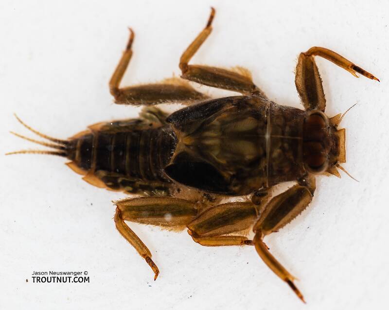
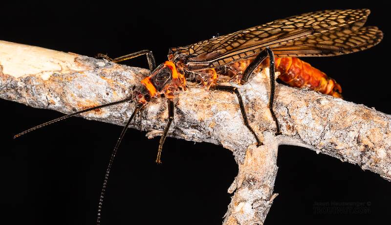
Pteronarcys californica
The giant Salmonflies of the Western mountains are legendary for their proclivity to elicit consistent dry-fly action and ferocious strikes.
Featured on the forum
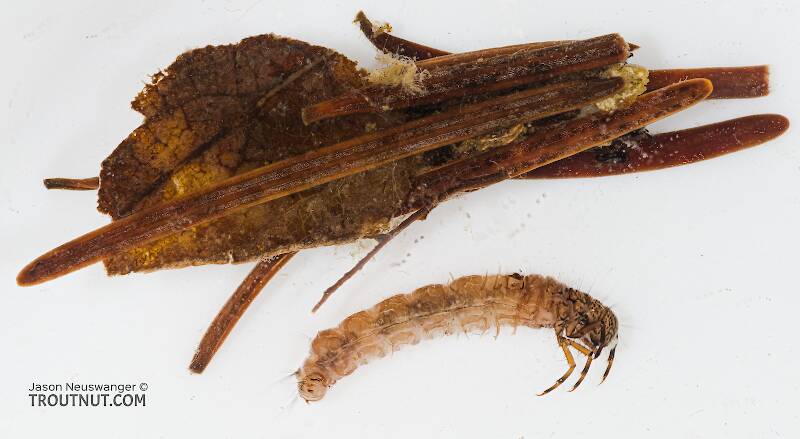
It's only barely visible in one of my pictures, but I confirmed under the microscope that this one has a prosternal horn and the antennae are mid-way between the eyes and front of the head capsule.
I'm calling this one Pycnopsyche, but it's a bit perplexing. It seems to key definitively to at least Couplet 8 of the Key to Genera of Limnephilidae Larvae. That narrows it down to three genera, and the case seems wrong for the other two. The case looks right for Pycnopsyche, and it fits one of the key characteristics: "Abdominal sternum II without chloride epithelium and abdominal segment IX with only single seta on each side of dorsal sclerite." However, the characteristic "metanotal sa1 sclerites not fused, although often contiguous" does not seem to fit well. Those sclerites sure look fused to me, although I can make out a thin groove in the touching halves in the anterior half under the microscope. Perhaps this is a regional variation.
The only species of Pycnopsyche documented in Washington state is Pycnopsyche guttifera, and the colors and markings around the head of this specimen seem to match very well a specimen of that species from Massachusetts on Bugguide. So I am placing it in that species for now.
Whatever species this is, I photographed another specimen of seemingly the same species from the same spot a couple months later.

Troutnut is a project started in 2003 by salmonid ecologist Jason "Troutnut" Neuswanger to help anglers and
fly tyers unabashedly embrace the entomological side of the sport. Learn more about Troutnut or
support the project for an enhanced experience here.
Identification: Key to Species of Drunella Nymphs, Couplet 3
Identification: Key to Species of Drunella Nymphs, Couplet 3
Adapted from Jacobus et al. (2014)
| Option 1 | Option 2 |
|---|---|
Forefemur with margins toothed 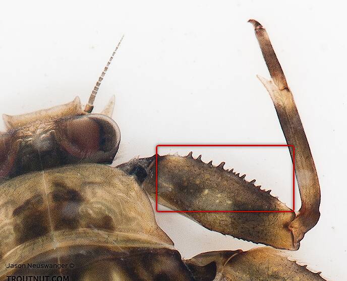
| Forefemur with margins smooth 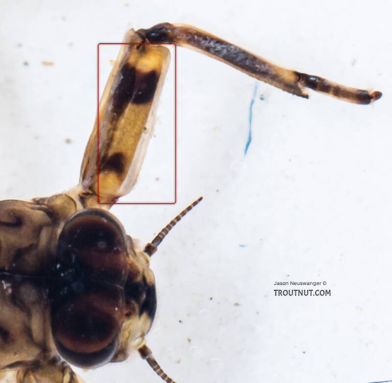
|
5 Example Specimens | 2 Example Specimens |
| Drunella doddsii | Drunella pelosa |
Adapted from Jacobus et al. (2014), Allen & Edmunds (1962), McCafferty et al (2017), and Jacobus LM, McCafferty WP (2004)
The current couplet is highlighted with darker colors and a icon, and couplets leading to this point have a icon.
Leads to Couplet 2:
- Western North American species
Couplet 2
Leads to Couplet 7:
- Eastern North American species
Couplet 7
Leads to Couplet 3:
- Abdominal sterna with prominent friction disc of setae
Couplet 3
Leads to Couplet 4:
- Abdominal sterna without prominent friction disc of setae (although felt-like coating of setae may be present)
Couplet 4
Couplet 3 (You are here)
Leads to Drunella doddsii:
- Forefemur with margins toothed
Leads to Drunella pelosa:
- Forefemur with margins smooth
Leads to Couplet 5:
- Forefemur with margins with prominent teeth
- Prosternum with no anterior projection
Couplet 5
Leads to Couplet 6:
- Forefemur with margins mostly smooth
- Prosternum with prominent anterior projection
Couplet 6
Leads to Drunella coloradensis:
- Abdominal terga with paired, narrow spines
Leads to Drunella flavilinea:
- Abdominal terga with paired, broadly triangular spines
Leads to Drunella spinifera:
- Abdominal terga 8 and 9 with paired spines more than twice the length of spines on preceding segments
Leads to Drunella grandis:
- Abdominal terga 8 and 9 with paired spines clearly less than twice the length of preceding pairs
Leads to Couplet 8:
- Abdomen without paired dorsal tubercles, but may have paired ridges
- Head smooth, without paired occipital tubercles (pictured yellow box)
Couplet 8
Leads to Couplet 10:
- Abdomen with paired dorsal tubercles, always well-developed on segments 5-7
- Head roughened, with paired occipital tubercles, or with small to large paired occipital tubercles
Couplet 10
Leads to Drunella lata:
- Frontoclypeal projections are short (approximately as long as their width at the base), not curved, and do not protrude anteriorly from the surface of the frontoclypeus (Funk et al. 2008)
- Femora with few or no long, fine setae on posterior margin (McCafferty et al 2017)
Leads to Couplet 11:
- Frontoclypeal projections are about twice as long as wide (extending to anywhere from just before the margin of the frontoclypeus to well beyond it), conical (or curved), and protrude anteriorly from the surface of the frontoclypeus (Funk et al. 2008)
- Femora with dense row of long, fine setae on posterior margin (McCafferty et al 2017)
Couplet 11
Leads to Drunella tuberculata:
- Has a row of long, hairlike setae along the margin of the clypeus
- More robust, less flattened general appearance
- Almost always has a pair of prominent, paired, stout, suboccipital spines
Leads to Drunella walkeri:
- Lacks a row of long, hairlike setae on the clypeus
- Usually a very flattened general appearance
- Sometimes has only small bumps, other times more prominent suboccipital spines
Leads to Drunella allegheniensis:
- Larvae have short, marginal, hairlike setae on the heads, legs, and tergites
- No long setae protruding dorsally from tergites 8 and 9
- Relatively many denticles on tarsal claws, and positioned along the length of the claw
Leads to Couplet 9:
- Marginal setae not well developed on the heads, legs, and tergites
- Long setae protrude dorsally from the anterior margins of tergites 8 and 9
- Relatively few denticles on tarsal claws, positioned basally
Couplet 9
Leads to Drunella longicornis:
- Foretibial projections long and distinctly curved
- Relatively uncommon; species status not 100 % certain
Leads to Couplet 12:
- Foretibial projections not developed as above
- Relatively widespread and common
Couplet 12
Leads to Drunella cornuta:
- Mature nymph 7.8-11.4 mm long (Funk et al. 2008)
- Median ocellar tubercle sharp and relatively long (red box)
- Lateral frontoclypeal projections usually semilunar (green boxes above)
- Middle and hind tibiae relatively long (Funk et al. 2008)
Leads to Drunella cornutella:
- Mature nymph 6.4-8.5 mm long (Funk et al. 2008)
- Median ocellar tubercle blunt to moderately sharp and relatively short
- Lateral frontoclypeal projections moderately curved to straight
- Middle and hind tibiae relatively short (Funk et al. 2008)
Start a Discussion of this Couplet
References
- Allen, R.K., and Edmunds, George F. Jr. 1962. A Revision of the Genus Ephemerella (Ephemeroptera, Ephemerellidae) V. The Subgenus Drunella in North America. Miscellaneous Publications of the Entomological Society of America 4: 147-179.
- Funk, D.H., Sweeney, B.W, and Jackson, J.K. 2008. A taxonomic reassessment of the Drunella lata (Morgan) species complex (Ephemeroptera:Ephemerellidae) in northeastern North America. Journal of the North American Benthological Society 27(3): 647-663.
- Jacobus, L. M., Wiersema, N.A., and Webb, J.M. 2014. Identification of Far Northern and Western North American Mayfly Larvae (Insecta: Ephemeroptera), North of Mexico; Version 2. Joint Aquatic Science meeting, Portland, OR. Unpublished workshop manual. 1-176.

