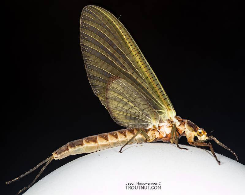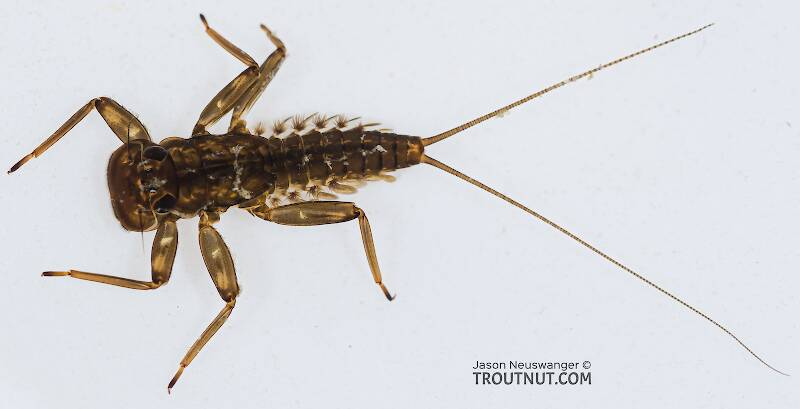
Hex Mayflies
Hexagenia limbata
The famous nocturnal Hex hatch of the Midwest (and a few other lucky locations) stirs to the surface mythically large brown trout that only touch streamers for the rest of the year.


Mayfly Species Apobaetis etowah
Species Range
Identification
Species ID from GBIFthe Global Biodiversity Information Facility
Source: Redescription of three species of Apobaetis Day, 1955 (Ephemeroptera: Baetidae)
Diagnosis. Male imago. 1) posterior margin of styliger plate triangular, area between unistyligers without emargination (Fig. 13 in Day 1955). Larva. 1) labrum rectangular, distal margin without shallow medial emargination, dorsal surface with three to four short and blunt setae medially near distal margin (Fig. 2 A); 2) lingua subquadrangular, with one medial protuberance (Fig. 2 D); 3) maxillary palp long, 1.6 × length of galea-lacinia, apex of segment II with constriction (Fig. 2 E); 4) labial palp segment II with pointed and apically directed distomedial projection, segment III rectangular, distal margin deeply concave (Fig. 2 F); 5) tarsal claws 0.85 × length of tarsus, without row of denticles (Fig. 3 A); 6) posterior margin of terga IV with triangular, pointed spines (Fig. 3 C).
Physical description
Most physical descriptions on Troutnut are direct or slightly edited quotes from the original scientific sources describing or updating the species, although there may be errors in copying them to this website. Such descriptions aren't always definitive, because species often turn out to be more variable than the original describers observed. In some cases, only a single specimen was described! However, they are useful starting points.
Male Spinner
Wing length: 4 mm
Abdominal tergites 2-6 of male imago semi-hyaline, white; marked with dots and dashes of red.
Head of male reddish brown; antennae very pale red-brown. Turbinate eyes very large, oval, light orange (alcoholic specimens); far apart at the anterior margins, closely approximated at the rear. Thorax reddish brown; postero-lateral margins of mesonotum narrowly pale yellowish; intersegmental areas of pleura, and an area anterior to wing root, yellowish. Legs whitish, coxae reddish brown. Wing hyaline, venation pale; main veins of anterior half of wing reddish brown at extreme base. A small faint whitish semi-opaque cloud in the stigmatic area; cross veins of this area few in number (about 4 to 6), simple, somewhat aslant; often with short horizontal vein traces between them. No intercalaries in first interspace; one or two, very short, in second space; other parts well developed.
Abdominal segments 2-6 white, semi-hyaline, a pair of small red submedian dots near the anterior margin of each tergite; another small red dot, or a pair of small dots close together, on each side, near middle of tergite, about halfway to the pleural fold; traces of geminate red mid-dorsal streaks, best developed on tergites 2, 3 and 6; and short red transverse dashes, one on each side of mid-dorsal line, near the posterior margin. Broken geminate blackish streaks mark the spiracular area. Tergites 7-10 reddish brown; sternites whitish or yellowish white. Forceps and tails white. Extending out between the bases of the forceps is a truncate ‘penis-cover,' which by reason of its length and shape seems unique in this group (see fig. 168).
The more numerous red dots and dashes on the tergites, the paler thorax, and the lack of ventral markings, distinguish this species from P. virile (now a synonym of Plauditus virilis).
Female Spinner
Head and pronotum of female flesh-colored; faint brownish markings on the vertex, and a brown transverse band across the pronotum. Remainder of thorax pale reddish brown; pale areas on mesonotum on each side of a brownish median stripe, and anterior to the scutellum; other large pale areas on the pleura. Dorsum of abdomen pale reddish, with traces of red markings as in the male; tracheae more or less outlined in black. Venter pale yellowish.
Description from GBIFthe Global Biodiversity Information Facility
Source: Redescription of three species of Apobaetis Day, 1955 (Ephemeroptera: Baetidae)
Redescription. Larva. Head: antenna with minute spines and fine, simple setae on apex of each segment. Frons with two keels. Labrum (Fig. 2 A): rectangular, broader than long; length about 0.6 × maximum width; distal margin without shallow medial emargination; ventral surface with robust spine-like setae on distolateral and distal margin; dorsal surface with three to four short and blunt setae medially near distal margin; dorsal surface with one row of long and thin setae medially, near distal margin. Right mandible (Fig. 2 B): incisors deeply cleft in two sets; outer and inner set of incisors with 3 and 2 – 3 denticles, respectively; prostheca slender, bifurcated at apex; margin between prostheca and mola concave; tuft of spine-like setae at base of mola absent; denticles of mola not constricted; lateral margin convex. Left mandible (Fig. 2 C): incisors deeply cleft in two sets; outer and inner set of incisors with 5 and 3 denticles, respectively; prostheca robust, bifid at middle, inner lobe slender, outer lobe robust; margin between prostheca and mola concave; tuft of spine-like setae at base of mola absent; subtriangular process wide; denticles of mola not constricted; lateral margin convex. Hypopharynx (Fig. 2 D): lingua subquadrangular, with one medial protuberance, and short apical tuft of setae, subequal than superlingua; superlingua not expanded, with short, fine, simple setae scattered over distal margin. Maxilla (Fig. 2 E): maxillary palp long, 1.6 × length of galea-lacinia; apex of segment II with constriction, similar to a third segment; maxillary palp with fine and simple setae scattered over surface. Labium (Fig. 2 F): glossa broad basally, narrowing slightly apically, subequal in length to paraglossa; inner margin with one row of blunt setae; apex with three short spine-like setae; outer margin with six spine-like setae; ventral surface covered with thin setae. Paraglossa curved inward; apex with two spine-like setae; outer margin with one row of ten robust spine-like setae; dorsal surface with one longitudinal row of seven robust spine-like setae near inner margin; ventral surface with one longitudinal row of five robust spine-like setae at middle. Labial palp with segment I 0.8 × length of segments II and III combined; segment I covered with micropores (not illustrated); segment II with pointed and apically directed distomedial projection, apex of distomedial projection slightly concave, outer margin and distomedial projection covered with fine, long and simple setae; inner margin bare; segment III rectangular, length 0.5 × width, covered with fine, long and simple setae on outer margin, ventral surface with five robust spine-like setae near outer margin, distal margin with one row of six robust spinelike setae, distal margin deeply concave. Thorax. Foreleg. Femur (Fig. 3 A): dorsally with row of 12 short concave and blunt setae (similar with Fig. 5 C in Cruz et al. 2018); apex with two concave and blunt setae; ventrally with row of elongated spine-like setae. Tibia. Dorsally bare; ventrally with one row of nine short spine-like setae. Patella-tibial suture present, apparently restricted to ventral margin (Fig. 3 B). Tarsus: bare dorsally; ventrally with one row of nine short spine-like setae. Tarsal claws 0.8 × length of tarsus, row of denticles absent. Abdomen: terga surface covered with scale-like triangular spines, micropores and short, fine and simple setae (not illustrated); posterior margin with triangular, pointed spines (Fig. 3 C). Gills as in Figure 1 in Day (1955). Paraproct (Fig. 3 D) with marginal spines, posterolateral extension with spines. Cerci with small lateral spines on all segments, paracercus without spines.
Start a Discussion of Apobaetis etowah
References
- Needham, James G., Jay R. Traver, and Yin-Chi Hsu. 1935. The Biology of Mayflies. Comstock Publishing Company, Inc.
Mayfly Species Apobaetis etowah
Species Range
Resources
- NatureServe
- Integrated Taxonomic Information System
- Global Biodiversity Information Facility
- Described by Traver (1935)

