Blue-winged Olives
Baetis
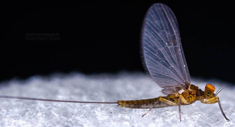
Tiny Baetis mayflies are perhaps the most commonly encountered and imitated by anglers on all American trout streams due to their great abundance, widespread distribution, and trout-friendly emergence habits.

- The prosternal horn is present.
- The mandible is clearly toothed, not formed into a uniform scraper blade.
- The seems to be only 2 major setae on the ventral edge of the hind femur.
- Chloride epithelia seem to be absent from the dorsal side of any abdominal segments.
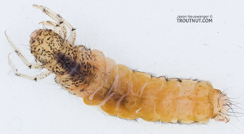
Based on these characteristics and the ones more easily visible from the pictures, this seems to be Grammotaulius. The key's description of the case is spot-on: "Case cylindrical, made of longitudinally arranged sedge or similar leaves," as is the description of the markings on the head, "Dorsum of head light brownish yellow with numerous discrete, small, dark spots." The spot pattern on the head is a very good match to figure 19.312 of Merritt R.W., Cummins, K.W., and Berg, M.B. (2019). The species ID is based on Grammotaulius betteni being the only species of this genus known in Washington state.

Stonefly Species Bolshecapnia spenceri (Little Snowflies)
Species Range
Physical description
Most physical descriptions on Troutnut are direct or slightly edited quotes from the original scientific sources describing or updating the species, although there may be errors in copying them to this website. Such descriptions aren't always definitive, because species often turn out to be more variable than the original describers observed. In some cases, only a single specimen was described! However, they are useful starting points.
Description from GBIFthe Global Biodiversity Information Facility
Source: A Review Of The Genus Bolshecapnia Ricker, 1965 (Plecoptera: Capniidae), And Recognition Of Two New Nearctic Capniid Genera
Male epiproct (n = 6). Length 524 - 543 µm, width at mid-length 224 - 250 µm, greatest width near base 295 - 300 µm. Sclerotized hooks arise subapically from either side of the median groove, and are bent sharply laterad, and extend beyond the lateral margins of the epiproct body (Figs. 25 - 29); tips of basolateral hooks extend forward for about 0.75 of the total epiproct length. Median groove wide near apex, narrowing gradually to the widest point near the epiproct base (Fig. 26). Small clumps of spongy appearing tissue located along lateral margins near base of hooks (Figs. 27 - 28). Base of epiproct body bearing a pair of dorsal ridges separated by terminus of median groove (Figs. 27, 29). Apex with a protruding membranous process (Figs. 29 - 30). Tergal process (n = 3). Absent, but tergum 9 covered with a broad band of short, thick setae (Figs. 27 - 28). Vesicle (n = 1). Length = 219 µm, basal width = 214 µm, median width = 252 µm. Process relatively wide, slightly wider near mid-length (Fig. 31 - 32). Ventral surface covered with thick setae.
Female subgenital plate (n = 3). This structure is an apically narrowed, tongue-shaped process, about twice as wide at mid-length as near the apical margin (Figs. 33 - 34); the structure extends beyond the anterior margin of sternum 9 (see fig. 169 in Baumann et al. 1977), and is hairless except for a few scattered long setae on the basal half. Several variations in the structure are shown in figs. 15 - 16 (Ricker 1965).
Start a Discussion of Bolshecapnia spenceri
References
- Merritt R.W., Cummins, K.W., and Berg, M.B. 2019. An Introduction to the Aquatic Insects of North America (Fifth Edition). Kendall/Hunt Publishing Company.
Stonefly Species Bolshecapnia spenceri (Little Snowflies)
Species Range
Common Names
Resources
- NatureServe
- Integrated Taxonomic Information System
- Global Biodiversity Information Facility
- Described by Ricker, W.E. (1965) New records and descriptions of Plecoptera (class Insecta). Journal of the Fisheries Research Board of Canada 22, 475–501.

