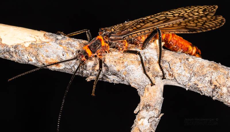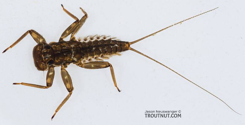
Salmonflies
Pteronarcys californica
The giant Salmonflies of the Western mountains are legendary for their proclivity to elicit consistent dry-fly action and ferocious strikes.


Caddisfly Species Palaeagapetus celsus (Microcaddisflies)
Species Range
Physical description
Most physical descriptions on Troutnut are direct or slightly edited quotes from the original scientific sources describing or updating the species, although there may be errors in copying them to this website. Such descriptions aren't always definitive, because species often turn out to be more variable than the original describers observed. In some cases, only a single specimen was described! However, they are useful starting points.
Description from GBIFthe Global Biodiversity Information Facility
Source: The genus Palaeagapetus Ulmer (Trichoptera, Hydroptilidae, Ptilocolepinae) in North America
Wings (A). Length of each forewing and hind wing 3.4 mm and 3.0 mm in male (3.1 – 3.6 mm, 2.6 – 3.2 mm, n = 6); 3.4 mm and 2.9 mm in female (3.0 – 3.6 mm and 2.7 – 3.0 mm, n = 9). Color and venation as in Palaeagapetus nearcticus. Lateral bulges of sternum V (7 B, F) round. Ventral process developed on segment VII in male (7 B) and segment VI of female (7 F). Male genitalia (Figs. 7 C – E). Segment IX (IX) short, anterolateral margins long, strongly projecting to anterior of segment VIII. Lateral appendages (la ap) of segment IX developed from mid-lateral region of genital capsule and directed caudad; thick and bilobed into dorsal and ventral branches at basal 1/3; dorsal branches (db) gently curved and tapered apically, each with slender process (sp) at basal 1/3 of mesal surface and many thick setae at apical half of dorsal surface, slender process directed mesocaudad with seta apically; ventral branches (vb) emerging near ventral bases of dorsal branches, directed ventrocaudad, each with 3 thick setae apically. Tergite X (tX) depressed dorsoventrally, curved dorsad apically in lateral view (7 C), semicircular in dorsal view (7 D). Inferior appendages (ia) each thick, short, twice as long as basal width, tapered at apical half with seta apically. Phallus (ph) short and broad, membranous with small forklike structure inside (7 C, E).
Female genitalia (Figs. 7 F, G). Segments I – VII each with sclerotized tergite and sternite, very setose, tergite VIII unpigmented. Segments IX – X very short, each segment about 1/2 as long as segment VIII, with somewhat developed cerci. Vaginal apparatus (7 G) slender, lateral projections undeveloped, lateral bands round.
Living Larvae. While examining live larvae under the microscope, JSW observed that the lateral processes of abdominal segments I – VIII do not resemble truncated fleshly tubercles of dead specimens as described by Flint (1962), but are actually much larger, membranous spheres. JSW also observed when a larva was prodded, to force it to exit its case, that the individual would sometimes turn 180 ° while staying completely inside its case. Then its head and thorax would emerge from the other end of its case and the larva would proceed to crawl away in the opposite direction (in respect to its previous position). The larva had the ability to use either end of its case as an opening for its head and thoracic legs, and the two ends of the case seemed to be rather similar in structure and function, similar in these ways to the larval behavior and case structure of distantly related Glossosoma spp. (Glossosomatidae) and Setodes spp. (Leptoceridae) (Wiggins 1996).
Start a Discussion of Palaeagapetus celsus
Caddisfly Species Palaeagapetus celsus (Microcaddisflies)
Species Range
Common Name
Resources
- NatureServe
- Integrated Taxonomic Information System
- Global Biodiversity Information Facility
- Described by Ross (1938)

