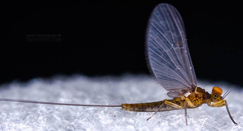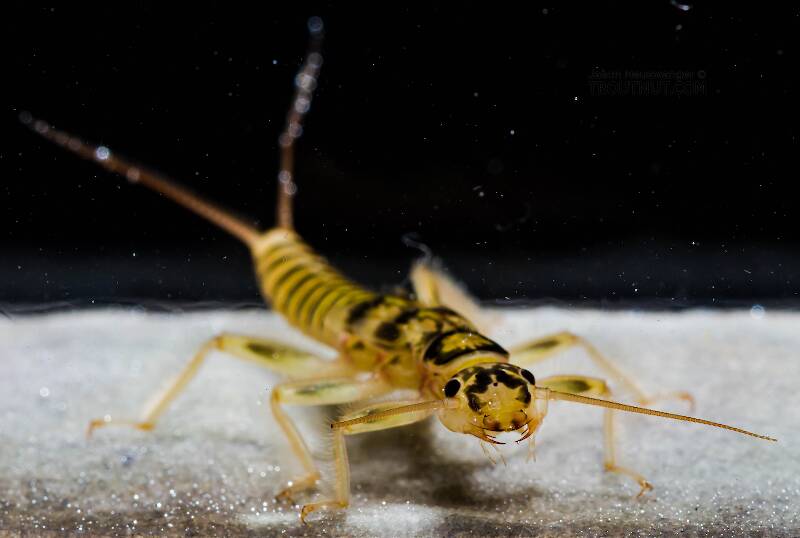
Blue-winged Olives
Baetis
Tiny Baetis mayflies are perhaps the most commonly encountered and imitated by anglers on all American trout streams due to their great abundance, widespread distribution, and trout-friendly emergence habits.


Stonefly Species Nemoura arctica (Tiny Winter Blacks)
Species Range
Identification
Species ID from GBIFthe Global Biodiversity Information Facility
Cercus. Highly variable. Adults of Nemoura arctica and Nemoura trispinosa have been previously differentiated by a combination of cercal characteristics (males) and body size and distribution (females) (Ricker 1952). Male cerci are sclerotized laterally and terminate typically in a pair of appressed spines that vary in length and degree of tapering (Figs. 1 – 16), plus a third unit that is highly variable and has been used for the past ca. 65 years to separate males of Nemoura arctica and Nemoura trispinosa (Ricker 1952). Lillehammer (1972 a) later illustrated cerci as either lacking (Nemoura arctica, his Fig. 4 b) or possessing a distinct (Nemoura trispinosa, his Fig. 4 a) spine. Ricker’s (1952, p. 36) key to Nemoura trispinosa males focused on “ … the outer edge of the cercus produced into a slender acute spine … may be forked once or twice at the tip ”. This feature is common in North America and shown here clearly for populations from Iowa (Fig. 1).
Physical description
Most physical descriptions on Troutnut are direct or slightly edited quotes from the original scientific sources describing or updating the species, although there may be errors in copying them to this website. Such descriptions aren't always definitive, because species often turn out to be more variable than the original describers observed. In some cases, only a single specimen was described! However, they are useful starting points.
Description from GBIFthe Global Biodiversity Information Facility
Source: Nearctic Nemoura Trispinosa Claassen, 1923 And Nemoura Rickeri Jewett, 1971 Are Junior Synonyms Of Holarctic Nemoura Species (Plecoptera: Nemouridae)
Epiproct. Males exhibit consistency with epiproct shape and characteristics across the Holarctic with only minor differences between individuals. In lateral aspect, the basal cushion occupies the anterior ca. ½ and is separated from the dorsal sclerite by smooth lateral areas (Figs. 17 – 24). The lateral areas vary in thickness but are consistently recurved slightly over the distal medial portion of the basal cushion. The dorsal sclerite appears scaly at high magnifications, especially at the distal tips (Figs. 41 – 52). The dorsal sclerite is open apically, exposing parallel, broad, hatchet-like apical prongs of the ventral sclerite (Figs. 25 – 40) and prominent, scaly, apical prongs positioned ca. perpendicular to the ridges (Figs. 41 – 52). The prongs terminate laterally bearing two short, thick, grooved spines (e. g. Figs. 42, 45, 49, 52).
Start a Discussion of Nemoura arctica
Stonefly Species Nemoura arctica (Tiny Winter Blacks)
Species Range
Common Names
Resources
- NatureServe
- Integrated Taxonomic Information System
- Global Biodiversity Information Facility
- Described by Esben-Petersen (1910) Bidrag til en fortegnelse over arktisk Neuropterfauna II. Tromsø Museums Årshefter 31/32, 82–86.

