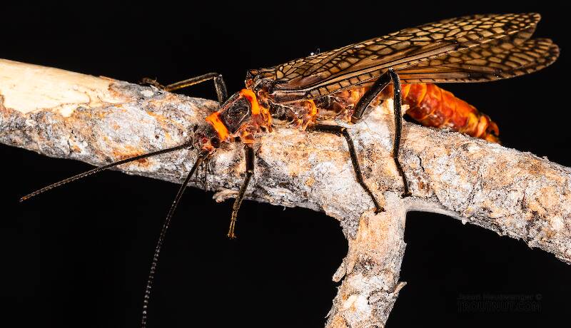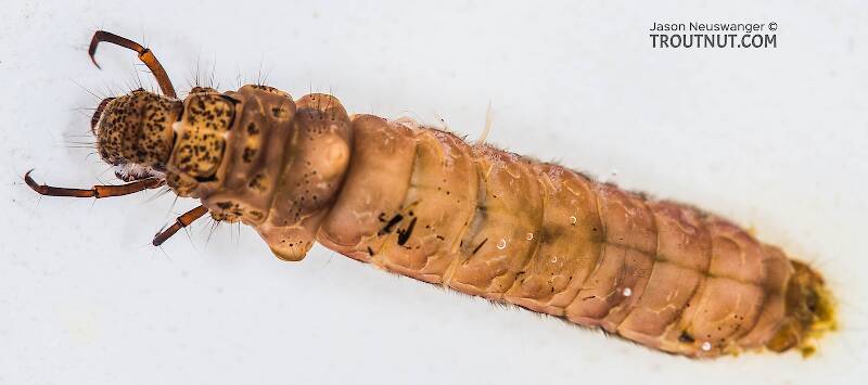
Salmonflies
Pteronarcys californica
The giant Salmonflies of the Western mountains are legendary for their proclivity to elicit consistent dry-fly action and ferocious strikes.


Stonefly Species Bolshecapnia gregsoni (Little Snowflies)
Species Range
Physical description
Most physical descriptions on Troutnut are direct or slightly edited quotes from the original scientific sources describing or updating the species, although there may be errors in copying them to this website. Such descriptions aren't always definitive, because species often turn out to be more variable than the original describers observed. In some cases, only a single specimen was described! However, they are useful starting points.
Description from GBIFthe Global Biodiversity Information Facility
Source: A Review Of The Genus Bolshecapnia Ricker, 1965 (Plecoptera: Capniidae), And Recognition Of Two New Nearctic Capniid Genera
Male epiproct (n = 3). Length 544 - 593 µm, width at mid-length 185 - 213 µm, greatest width near base 147 - 253 µm. A pair of curved, acute, sclerotized hooks arise from either side of the median groove at about half the distance between the epiproct apex and the base of the spongy area from either side of the median groove. The hooks extend slightly beyond lateral margins of the epiproct body (Figs. 1 - 3), and their tips reach to about 0.8 of the epiproct length. Median groove wide near apex and narrowed near dorsobasal knobs (Fig. 4). Median groove divides a pair of spongy-appearing clumps of tissue near hooks (Fig. 2). Apex without a protruding membranous process; dorsobasal humps low, smooth and not outlined by a prominent posterodorsal ridge (Fig. 2 - 3). Paraprocts with a hairy, plate-like basal area and slender apices. Tergal process (n = 3). Absent, but tergum 9 bears a median patch of short, thick setae (Fig. 1) and lacks a median anterior notch. However, tergum 10 bears a median anterior notch filled with membranous tissues projecting into the notch from tergum 9 (Fig. 7). Vesicle (n = 1). Length 305 µm, width at mid-length 311 µm, basal stalk short and 168 µm wide. Surface entirely covered with thick setae except for the short stalk that extends under the basal roll of tissue (Fig. 4).
Female subgenital plate (n = 2). This structure is a triangular plate that extends beyond the anterior margin of sternum 9 (see fig. 10 in Ricker 1965, fig. 173 in Baumann et al. 1977, and fig. 3.12 in Stewart & Oswood 2006). The images we present show an almost triangular plate, rounded and glabrous at the apex with convergent lateral margins (Figs. 5 - 6). The transverse striations observed on the subgenital plate in Fig. 6 may be an artifact of dehydration.
Start a Discussion of Bolshecapnia gregsoni
Stonefly Species Bolshecapnia gregsoni (Little Snowflies)
Species Range
Common Names
Resources
- NatureServe
- Integrated Taxonomic Information System
- Global Biodiversity Information Facility
- Described by Ricker, W.E. (1965) New records and descriptions of Plecoptera (class Insecta). Journal of the Fisheries Research Board of Canada 22, 475–501.

